Categories
- Anatomical Models/Charts -
- -Alternative
- -Brain/Head
- -Budget Items
- -Dental
- -Digestive
- -Ear/Nose/Throat
- -Exercise/Diet/Health
- -Extremities
- -Eye
- -Female
- -For Kids
- -General Anatomy
- -Heart
- -Immune System
- -Joint
- -Male
- -Muscle
- -Respiratory
- -Skeleton
- -Skin
- -Skull
- -Spanish
- -Stands & Displays
- -Torso
- -Training Aids
- -Urinary
- -Vascular
- -Vertebrae/Spine
- Halloween Products -
- Charts
- Models
- Reference
- Study Guides
- Gifts
- Stands & Displays

")
Anatomical Models/Charts


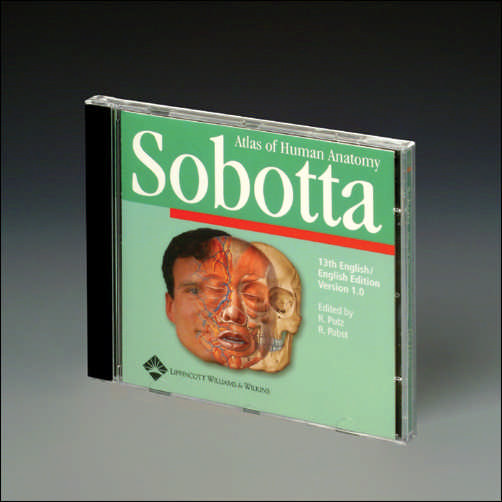
SOBOTTA ATLAS OF HUMAN ANATOMY, VERSION 1.5
Contains over 1500 illustrations and tables from the 13th English edition of the Sobotta Atlas. Including detailed drawings made from dissections, cross-sections and classical anatomy and reproductions of imaging techniques. 13th Edition in English, CD-ROM for Windows.
ISBN 10: 0-7817-4054-1 ISBN 13: 978-0-7817-4054-8
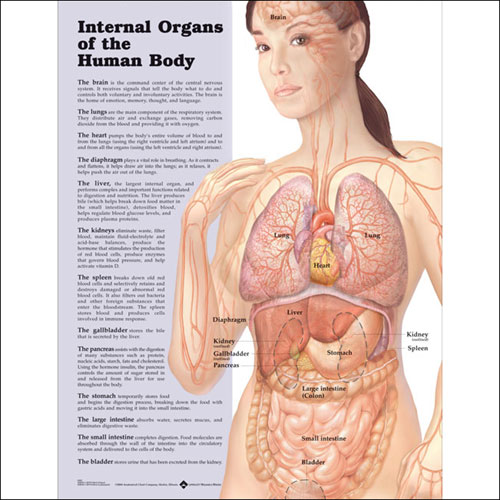
INTERNAL ORGANS OF THE HUMAN BODY – FLEXIBLE LAMINATION
This chart provides a simple and easy-to-understand overview of the location and functions of the major internal organs of the body, including: heart, lungs, stomach, kidney, diaphragm, spleen, liver, pancreas, large and small intestine, gallbladder, bladder and brain. Perfect for patients and students. Size is 20" W by 26" H. For more information about chart mounting styles, click here.
Paper: ISBN 10: 1-58779-828-X ISBN 13: 978-1-58779-828-3
Laminated: ISBN 10: 1-58779-829-8 ISBN 13: 978-1-58779-829-0
INTERNAL ORGANS OF THE HUMAN BODY – FLEXIBLE LAMINATION
This chart provides a simple and easy-t...
$11.95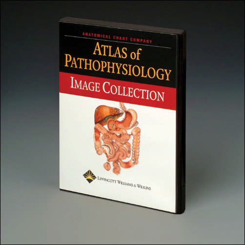
ATLAS OF PATHOPHYSIOLOGY IMAGE COLLECTION
Designed to complement the Atlas of Pathophysiology, this CD-ROM image collection contains approximately 300 images from the atlas for use in lecture presentations. Images can be imported into a PowerPoint presentation, projected directly from the program onto a screen, or printed and copied onto overhead transparencies. Images are available with callouts and leader lines already in place and are available in both JPG and PDF formats. The table of contents is organized by body system to help the reader locate images quickly and easily. Users can conveniently search for the desired pathophysiology, and thumbnail images allow instructors to quickly identify the image that best suits their needs. CD-ROM for Windows.
ISBN 10: 1-58255-263-0 ISBN 13: 978-1-58255-263-7
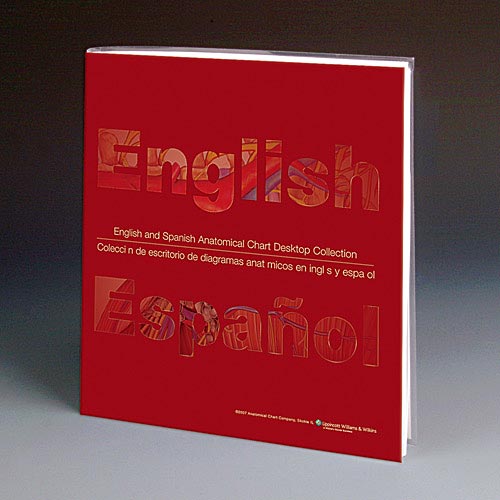
ENGLISH/SPANISH ANATOMICAL CHART DESKTOP COLLECTION
This full color book includes 34 of our most popular anatomy and pathology topics presented in both English and Spanish (68 charts total), with emphasis on those disorders that may often affect the Hispanic/Latino population.
Handy for learning terminology, this book is also very handy in bilingual patient counseling situations. The covered spiral binding allows the book to lay flat on desk with the chart in English on the left hand side, next to the same chart in Spanish on the right.
Printed in vivid lifelike colors on high-quality paper, the anatomical charts show the human body in a format that provides a clear and visual understanding of human anatomy, physiology and diseases. Medical terminology and easy to understand supporting text is printed directly on each chart so you never have to refer to a separate key card or manual. Size: 10" x 12".
$46.95
ENGLISH/SPANISH ANATOMICAL CHART DESKTOP COLLECTION
This full color book includes 34 of our...
$46.95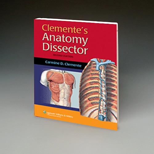
CLEMENTE'S ANATOMY DISSECTOR
A comprehensive lab manual of anatomical dissection referenced to current editions of major medical atlases. Organized into 32 chapters, each focuses on small discrete areas such as the posterior triangle of the neck, and dorsum of the hand. Provides in-depth, detailed explanations for each dissection. Each chapter also stands alone, which allows instructors to adapt the material to their preferred dissection sequence, and students to self-teach and review. Features correlation of the surface anatomy to anatomical structures revealed in the dissections, a highlight for clinical understanding. Dissection instructions are boxed in red to create clear distinction from the explanatory material. 311 illustrations, 400 pages, soft cover, spiral bound.
ISBN 10: 0-7817-3290-5 ISBN 13: 978-0-7817-3290-1
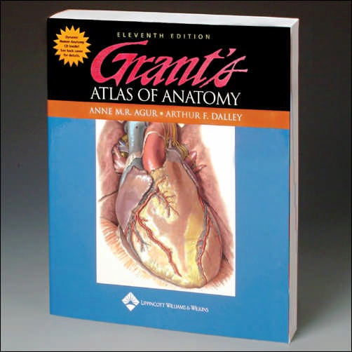
GRANT'S ATLAS OF ANATOMY 11TH EDITION
Since 1943 this classic has been providing students with accurate anatomical images presented in an effective easy to understand and clinically relevant manner. 1600 illustrations. 848 pages. Soft cover.
ISBN 10: 0-7817-4255-2 ISBN 13: 978-0-7817-4255-9
New Products For June - Anatomical Models/Charts


More Information
Bestsellers
Newsletter
LATEST PRODUCTS

Posabe Life-size pair of Harvey skele...
-

This is the aged version of the Posab...
EXTRAS
GET IN TOUCH
Find Us On Map
+1 269-646-9597

© Copyright 2025 by SkeletonandMore.com